Illustrate/sketch/printout of
photomicrographs (b/w) of all the tissue sections listed below. Label all
photomicrographs with the given description for each tissue section.
Objective: Differentiate between
the different types of epithelium; whether they are keratinized, stratified,
and whether they are ciliated or contain microvilli.
________________________________________________________________________________________
1. Simple squamous epithelium (isolated) – Label
Cell Membrane;ICS;Nucleus
________________________________________________________________________________________
2. Stratified squamous epithelium
non-keratinized from the vagina. The portion lining the lumen contains the
epithelium. Identify the types of cells
that make up this lining. The innermost
layers of cells are cuboidal,
but towards the surface they become squamous. Remember that in stratified epithelia,
the outer layer determines the classification of the epithelium. Below the lining of the epithelial cells lies
the region of connective tissue.
________________________________________________________________________________________
3. Stratified squamous from the esophagus
________________________________________________________________________________________
4. Simple cuboidal epithelium surrounding thyroid
follicles
________________________________________________________________________________________
5. Simple cuboidal to simple columnar in kidney. Look for a large Y-shaped region. This is the urinary space of the minor
calyx. Thin tubules enter/drain into
this space, having radiated through the light pink medulla region from the darker
red, cortex region. Microscopically, the
medulla contains tubules with cuboidal (cube-shaped, with a squarish profile)
or short columnar (taller than it is wide) epithelium. You will also find some very narrow tubules
with squamous (flattened cells) epithelial walls. The cortex has glomeruli with squamous
epithelium, and tubules with cuboidal epithelium.
________________________________________________________________________________________
6. Simple columnar epithelium of the pyloric
stomach. Label the unique cell types
that are present in the tissue section.
________________________________________________________________________________________
7. Pseudostratified columnar ciliated epithelium
in the respiratory passages (trachea).
Note that this is a slice of a tube.
Look at the inner lining to find the epithelial cells. These are pseudo-stratified (falsely layered)
because the nuclei are at different levels and the cells are of different
heights, but all cells sit on the basement membrane (which is not
obvious in this slide). Thus, there is
only a single layer of different-sized cells.
These epithelial cells are ciliated, and this differs from the
brush border of the gut epithelial cells.
Make sure you can tell the difference between cilia and microvilli of
the brush border. You will also see
goblet cells here. These are the
flask-shaped cells sitting on the outer 2/3 of the epithelium. The base of the goblet cell also sits on the
basement membrane. What is the purpose
of the cilia?
________________________________________________________________________________________
8. Transitional epithelium
of the urinary bladder. The bladder has a folded lumen (central space). This lumen is lined by transitional
epithelium. Note the layers of
epithelial cells. The outer layer of transitional epithelial cells (sometimes
called umbrella cells) has an outer lining of dark red material. This dark lining is formed by the
invagination of the membranes to accommodate stretching of the cell when the
lumen is expanded (when the bladder fills).
________________________________________________________________________________________
9. Stratified squamous epithelium
keratinized palm) . Compare this with
the stratified squamous epithelium of the vagina.
________________________________________________________________________________________
10. Tongue. The upper surface has papillae, which
are projections of the stratified squamous epithelium which create
friction. The papillae are
keratinized. This means that the
epithelial cells have died and produced a non-cellular layer of keratin on the
surface. Keratinized epithelium lines
the body surface for protection. For now
note the intact cells under the keratinized layer.
Spikes are false
intercellular bridges. These do not
connect the cytoplasm of adjacent cells, but are really sites of cell membrane
(junctions, desmosomes) that connect the adjacent epithelial cells. When the material was prepared, there was
shrinkage, and the cytoplasm pulled away from the next cell, leaving only the
strong desmosomal attachment, creating this appearance. “Stratified squamous” means that the
uppermost cells are squamous in appearance, and stratified means there is more
than one layer of cells.
________________________________________________________________________________________
11. Mitochondria from liver. Look for the little dark dots which are the
mitochondria. Identify the nuclei and
nucleoli.
________________________________________________________________________________________
12. Golgi Apparatus. In the electron microscope,
this is seen as a system of parallel membranes arranged around the
nucleus. It is seen as black splotches
in the cytoplasm of large round nerve cells.
These nerve cells gave a large, light nucleus with a dark dot
(nucleolus).
________________________________________________________________________________________
13. Scalp. The skin consists of stratified squamous
epithelium, keratinized. The basal layer
is cuboidal, but becomes squamous in the upper layers. The cells die and form keratin, which are
layers of protein that slough off regularly.
Identify the hair follicles, sebaceous (oil) glands, sweat
glands and smooth muscles making up the arrector pili muscle.
________________________________________________________________________________________
14. Gut epithelium / Small
intestine (duodenum) simple columnar cells. Identify finger-like
processes (villi). The lining of these villi contains a simple columnar
epithelium with a striated or brush border (the electron
micrograph). The brush border can be
seen as a thin amorphous pink layer at the top of the epithelial cells. In most preparations some cells are different
from the simple columnar epithelium.
These appear as dark shadows or clear spots on the luminal surface of
the epithelium as you focus up and down.
These are the goblet cells, which are unicellular mucous
secreting cells. .
________________________________________________________________________________________
15. Parotid gland. Identify the secreting portion
and the ducts. Most of the gland is make
up of serous secreting alveoli (sacs of cells), which have characteristic
round, rather light, central nuclei and pink cytoplasm. The ducts look like necklaces, with the cells
being cuboidal, stratified cuboidal, to columnar. Identify the large ducts in the spaces
between lobules – some of these are stratified cuboidal.


 Neurofibrillary Tangles associated with Alzheimer's
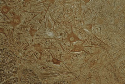
 Congo red histology stain is used to stain amyloid.


