THIS IS TO BE DONE INDIVIDUALLY.
NAME: ___________________________
· Always begin examining microscope slides with which power objective? ____________
· The lens that is within the eyepiece of the light microscope is called the: ____________
·What must be done to a specimen to increase the contrast of the structures viewed? ____________
· The wheel under the stage that adjusts the amount of light is called the: ____________
· At high power, always use which adjustment knob to focus the image? ____________
· To focus a specimen, it is best to start with which objective: ____________
· Which objective provides the greatest depth of field? ____________
·The scanning, low, and high power objectives are mounted on the: ____________
If the ocular of a microscope is 10X and the objective is set at 100X, then what is the total magnification of the microscope? ____________
If the ocular of a microscope is 10X and the objective is set at 100X, then what is the total magnification of the microscope? ____________
1. This ____ (organelle) functions in cellular respiration.
2. The ____ (organelle) functions to package and deliver proteins.
3. Cell organelles are located within the ____ of the cell.
4. The endoplasmic reticulum functions to____________________
5. Genetic material is contained within the ___ of the cell.
6. This organelle is responsible for destroying worn-out cell parts___
7. The _____ controls what enters and leaves the cell.
8. The rough endoplasmic reticulum has ____ located on it.
9. Located within the nucleus, it is responsible for producing ribosomes:
10. Which structure is directly responsible for the formation of proteins within the cell.
Pre-test on Mitosis
· What is mitosis?
· Beginning with mitosis, what are the other parts of the cell cycle in order in which they occur?
· What occurs in the cell during the G2 phase?
· Beginning with interphase, what are the phases of mitosis in order in which they occur?
· In which phase do the chromosomes align on a plane in the center of the cell?
· What occurs during anaphase?
· What is the part of the chromosome that joins the two chromatids?
· What is the structure in cells that divides the cytoplasm into two cells.
· What is a blastula?
· What is the term that describes a nucleus with two of each type of chromosome?
A quiz on this assignment will be given on Friday. DO YOUR HONEST BEST!
Alkaline Phosphatase
Bielschowsky Stain


 Neurofibrillary Tangles associated with Alzheimer's
Cajal Stain
Congo Red
Giemsa Stain

STAINS (Study in advance)
Aldehyde Fuchsin- Used to stain pancreatic islet beta cell granules.
- This stains elastic fibers purple/black.
Alician Blue
- Stain mucins and mucosubstances blue.
- Copper in the histology stain is what ultimately is responsible for the blue color.
- Nuclei will stain pink/red.
- Cytoplasm stains a lighter pink.
- Mucins stain blue.
Alkaline Phosphatase

- Stain endothelial cells.
- Sites of alkaline phophatase activity will appear red.
- Nuclei will stain blue.
Bielschowsky Stain
- Silver is used in this histology staining process.
- This histology stain shows reticular fibers.
- This histology stain is also used for showing neurofibrillary tangles and senile plaques.
- Neurofibrillary tangles and senile plaques will stain black.
 Neurofibrillary Tangles associated with Alzheimer's
Cajal Stain

- This histology stain is used on nervous tissue
Congo Red
Congo red histology stain is used to stain amyloid.
- Amyloid will stain orange/red.
Giemsa Stain

- This is a histology stain for peripheral blood smears and bone marrow.
- It is also used to visualize parasites and malaria.
- This is a Romanowski type stain.
- Methylene blue and eosin are used.
- Erythrocytes stain pink/red.
- Platelets and leukocytes stain blue.
This micrograph depicts the appearance of a well-stained slide using the Giemsa staining technique.
Note that the acidic components of the cellular constituents such as the cytoplasm and chromatin, pick up the basic methylene blue azure compliments of the Giemsa stain, which reveals the characteristic blue
coloration of this stain.
Golgi Stain
Periodic Acid-Schiff (PAS)


Note that the acidic components of the cellular constituents such as the cytoplasm and chromatin, pick up the basic methylene blue azure compliments of the Giemsa stain, which reveals the characteristic blue
coloration of this stain.
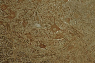
Golgi Stain
- This histology stain will stain neurons.
H&E

- This is a standard histology stain.
- "H&E" stand for hematoxylin and eosin.
- Hematoxylin and eosin stain is used for routine tissue preparation frequently.
- This is the most often used combination in the histology lab for general purpose staining.
- Hematoxylin can be thought of as a basic dye.
- It binds to acidic structures, staining them blue to purple.
- It will bind and stain nucleic acids.
- Therefore, the nucleus stains blue.
- Eosin is an acid aniline dye.
- It will bind to and stain basic structures (or negatively charged structures), such as cationic amino groups on proteins.
- It stains them pink.
- Cytoplasm, muscle, connective tissue, colloid, red blood cells and decalcified bone matrix all stain pink to pink/orange/red with eosin.
- With an H&E stain, mucus and cartilage will stain a light blue color.
Iron Hematoxylin
- 
- stain nuclei bluish/black
- one of a number of stains that allow one to make a permanent stained slide for detecting and quantitating parasitic organisms
Oil Red O

- This is a histology stain used for lipids.
- Lipids will stain red.
- Nuclei will stain blue/black

Papanicolaou Stain
- This histology stain is used mainly on exfoliated cytological specimens.
- Cells in smear preparations can be stained with Pap staining.
- Gynecological smears (Pap smears), sputum, urine, cerebrospinal fluid, abdominal fluid, pleural fluid, synovial fluid, semminal fluid and fine needle aspiration samples can all be stained with a Pap stain.
- This staining technique involving five dyes in three solutions.
Periodic Acid-Schiff (PAS)
- This histology stain is particularly useful for staining glycogen and other carbohydrates, but is useful for many things.
- It is often used to show glomeruli, basement membranes, and glycogen in the liver.
- PAS stains glycogen, mucin, mucoprotein, and glycoproteins magenta.
- The nuclei will stain blue.
- Collagen will stain pink.

Prussian Blue
- used to stain iron (ferric iron and ferritin).
- demonstrates the blue granules of hemosiderin in hepatocytes and Kupffer cells.- Hemochromatosis
Safranin O

- This histology stain will stain mucin, cartilage and mast cells.
- It stains them orange/red.
- Safranin O is sometimes used as a counterstain.


No comments:
Post a Comment