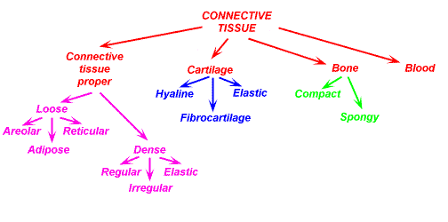
Of the four basic tissue types (epithelium, connective tissue, muscle and nervous tissue), connective tissue is the most diverse. Connective tissue can be found all over the body. Connective tissue cells are capable of reproducing, however not as fast as epithelial cells. Embryologically, connective tissue develops from mesenchyme.
Function of Connective Tissue
Connective tissues connects and holds tissues and organs to one another, forms a framework, provides support, stores fat, transports things, protects, and aids in tissue repair.
Histology of Connective Tissue
Connective tissue consists of cells and extracellular fibers in a ground substance and tissue fluid. The cells within connective are not tightly packed together. On a histology slide, it can be seen that there is generally abundant extracellular space in connective tissue.
Within connective tissue, the cells and fibers are scattered within the ground substance. The ground substance is amorphous material composed of proteoglycans and glycosaminoglycans. Hyaluronic acid is a glycosaminoglycan. Dermatan sulfate, chondroitin sulfate, and keratan sulfate are also
glycosaminoglycans.
Most of the connective tissues in the body have a good vascular supply.
Cell Types in Connective Tissue
Numerous cell types are found in connective tissue. Fibroblasts, histiocytes, plasma cells, and mast cells are cell types that are found in loose connective tissue.
Fibroblast
A fibroblast is a connective tissue cell derived from mesenchyme. Fibroblasts produce collagen. The fibroblast also produces the ground substance in connective tissue.
Histiocyte
The histiocyte is a connective tissue macrophage. Macrophages are mononuclear phagocytes, derived from a monocyte. A macrophage is a phagocytic tissue cell of the reticuloendothelial system that may be fixed or freely motile. Many tissues have resident (fixed) macrophages. Fixed macrophages are given a unique name, depending on the tissue that they are located in. In connective tissue, the fixed macrophage is a histiocyte. The function of histiocytes is for the protection of the body against infection and other harmful things.

Plasma cells are derived from B lymphocytes.
Mast Cell

A mast cell is a large cell seen in connective tissue. A mast cell has basophilic granules containing histamine and heparin which mediate allergic reactions. Mast cells also secrete SRS-A (slow reacting substance of anaphylaxis and ECF-A (eosinophilic chemotactic factor of anaphylaxis).
Fibers in Connective Tissue


There are three types of fibers found in connective tissue: collagen fibers, elastic fibers, and reticular fibers.
Collagen fibers are the most abundant fiber type in connective tissue.
Classification of Connective Tissue


Connective tissue can be sub-classified into connective tissue proper, specialized connective tissue and embryonic connective tissue.
Connective Tissue Proper

Connective tissue proper consists of loose irregular connective tissue and dense connective tissue. Dense connective tissue is subdivided into dense regular connective tissue and dense irregular connective tissue.


Loose Irregular Connective Tissue
 Loose irregular connective tissue is areolar tissue.
Dense Regular Connective Tissue
 Dense regular connective tissue comprises tendons and ligaments.
Dense Irregular Connective Tissue
Dense irregular connective tissue is seen in the dermis.
Specialized Connective Tissue
Specialized connective tissue includes cartilage, bone, adipose tissue, blood and hemopoietic tissue, and lymphatic tissue.
Cartilage
Bone
Adipose Tissue
Brown adipose tissue is multilocular adipose tissue. Brown adipose tissue is present during fetal development and then decreases after birth. White adipose tissue is unilocular adipose tissue. White adipose tissue persists into adulthood.

Blood and Hemopoietic Tissue
Lymphatic Tissue
Embryonic Connective Tissue
Embryonic connective tissue includes mesenchyme and mucous connective tissue. Mesenchyme is embryonic connective tissue. It is an undifferentiated tissue found in the embryo. Mucous connective tissue is a type of embryonic connective tissue; it is a subset of mesenchyme. Wharton's jelly is mucous connective tissue.
Histotechniques
Collagen fibers stain green with Masson's trichrome stain. Collagen fibers can be differentiated from other fibers by staining with Masson's trichrome stain. A trichrome stain is a mixture of three dyes.
Gomori's trichrome will stain connective tissue green, cytoplasm red, and nuclei gray blue black.
Verhoeff Elastic stain stains elastic fibers blue/black. Collagen stains pink/red.
KEY POINTERS FOR REVIEW:
- Of the four basic tissue types (epithelium, connective tissue, muscle and nervous tissue), connective tissue is the most diverse. Blood, bone, tendon, and intervertebral discs are all composed of connective tissue. The myometrium is the muscular layer of the uterus. Thus, the myometrium is composed of muscle tissue.
- There is connective tissue in the heart. The blood in the heart, for example, is composed of connective tissue. The pericardium is also composed of connective tissue. However, the primary tissue composing the heart is cardiac muscle.
- There are three types of fibers found in connective tissue: collagen fibers, elastic fibers, and reticular fibers. Collagen fibers are the most abundant fiber type in connective tissue.
- Myofibroblasts contain properties of both fibroblasts and smooth muscle cells.
- Fibroblasts, histiocytes, plasma cells, and mast cells are routinely seen in loose connective tissue. Fibroblasts produce collagen. The fibroblast also produces the ground substance in connective tissue. Myofibroblasts contain properties of both fibroblasts and smooth muscle cells. The histiocyte is a connective tissue macrophage. Plasma cells are derived from B lymphocytes. Mast cells secrete histamine. Mast cells also secrete heparin, SRS-A (slow reacting substance of anaphylaxis, ECF-A (eosinophilic chemotactic factor of anaphylaxis.
- Macrophages are mononuclear phagocytes. Many tissues have resident (fixed) macrophages. Fixed macrophages are given a unique name, depending on the tissue that they are located in. Kupffer cells are the hepatic macrophages. Histiocytes are macrophages seen in connective tissue. Dust cells are alveolar macrophage found in the respiratory tract. Langerhans cells are macrophages seen in the skin. Microglia are the central nervous system macrophages.
- Connective tissue can be sub-classified into connective tissue proper, specialized connective tissue and embryonic connective tissue.
- Connective tissue proper consists of loose irregular connective tissue and dense connective tissue (regular and irregular).
- Specialized connective tissue includes cartilage, bone, adipose tissue, blood and hemopoietic tissue, and lymphatic tissue.
- Embryonic connective tissue includes mesenchyme and mucous connective tissue.
- Mesenchyme is embryonic connective tissue. It is an undifferentiated tissue found in the embryo. Mucous connective tissue is a type of embryonic connective tissue; it is a subset of mesenchyme. Wharton's jelly is mucous connective tissue. Loose irregular connective tissue is areolar tissue. Dense irregular connective tissue is seen in the dermis. Dense regular connective tissue comprises tendons and ligaments.
- Brown adipose tissue is multilocular adipose tissue. This is present during fetal development and then decreases after birth.
- White adipose tissue is unilocular adipose tissue. This type of tissue persists into adulthood.
- A peripheral blood smear would be best visualized with Wright's stain. Hematoxylin and eosin stain is the most commonly used tissue stain for routine histological examination. Lipids are best displayed with a sudan stain. Silver impregnation, such as with a reticular stain, can be used to visualize reticular fibers. Collagen fibers can be differentiated from other fibers by staining with Masson's trichrome stain.
- Connective tissue develops from mesenchyme.
- Within connective tissue, the cells and fibers are embedded in the ground substance. The ground substance is amorphous material. It is composed of proteoglycans and glycosaminoglycans. Hyaluronic acid and chondroitin sulfate are glycosaminoglycans.
- Fibroblasts produce collagen. The fibroblast also produces the ground substance in connective tissue.
- Plasma cells are derived from B lymphocytes.
- Connective tissue consists of cells and extracellular fibers in a ground substance and tissue fluid. There is generally abundant extracellular space in connective tissue


