Thursday, December 8, 2016
Connective tissue Prelim Exam Requirement:
INDIVIDUALLY Submit a transcription of this video on connective tissue: on December 16, 2016
Tuesday, December 6, 2016
Connective tissue
1. Remember that all connective tissue contains
- -cells
- -fibers
- -collagen
- -elastic
- -reticular
- -ground substance (extracellular matrix)
- -fetal mesenchyme
- -mucoid connective tissue
- -loose, areolar fibrous CT (e.g., mesentery)
- -dense, irregular fibrous CT
- -dense, regular fibrous CT
- -elastic CT
- -reticular CT
- -adipose CT
- -white (unilocular) fat
- -brown (multilocular) fat
- -fibroblasts
- -adipocytes
- -macrophage
- -mast cell
- -plasma cell
- -leukocytes
6. List the general functions of connective tissues
7. Name the germ layer(s) from which connective tissue cells derive and the embryonic tissues containing undifferentiated connective tissue cells
9. Describe adipose tissue as a connective tissue in terms of its cells, fibers, and ground substance.
10. Compare the three types of cartilage in terms of:
- Type, amount, and arrangement of cells, fibers, and ground substance
- Location in the body
- Histogenesis
- Function
11. Compare osteocytes with osteoblasts in terms of their shape, filopodia, amount of RER, location, and rate of matrix synthesis.
SUBMIT ANSWERS PER GROUPS OF 8 MEMBERS EACH TO apbautista@ceu.edu.ph before 2 pm today (12-7-16)
Additional points will be given to:
1st - 20 pts to CP and 5 pts to the quiz on Friday
2nd - 15 pts
3rd - 10 points
Post lab discussion will be on Friday (12-9-16) and a short quiz will be given on connective tissue and epithelial tissue (30 pts)
PRELIM EXAM WILL BE ON FRIDAY NEXT WEEK (12-16-16)
Tuesday, November 22, 2016
Lab Activity Sheet: Epithelium, Skin and Glands
Illustrate/sketch/printout of
photomicrographs (b/w) of all the tissue sections listed below. Label all
photomicrographs with the given description for each tissue section.
Objective: Differentiate between
the different types of epithelium; whether they are keratinized, stratified,
and whether they are ciliated or contain microvilli.
________________________________________________________________________________________
1. Simple squamous epithelium (isolated) – Label
Cell Membrane;ICS;Nucleus
________________________________________________________________________________________
2. Stratified squamous epithelium
non-keratinized from the vagina. The portion lining the lumen contains the
epithelium. Identify the types of cells
that make up this lining. The innermost
layers of cells are cuboidal,
but towards the surface they become squamous. Remember that in stratified epithelia,
the outer layer determines the classification of the epithelium. Below the lining of the epithelial cells lies
the region of connective tissue.
________________________________________________________________________________________
3. Stratified squamous from the esophagus
________________________________________________________________________________________
4. Simple cuboidal epithelium surrounding thyroid
follicles
________________________________________________________________________________________
5. Simple cuboidal to simple columnar in kidney. Look for a large Y-shaped region. This is the urinary space of the minor
calyx. Thin tubules enter/drain into
this space, having radiated through the light pink medulla region from the darker
red, cortex region. Microscopically, the
medulla contains tubules with cuboidal (cube-shaped, with a squarish profile)
or short columnar (taller than it is wide) epithelium. You will also find some very narrow tubules
with squamous (flattened cells) epithelial walls. The cortex has glomeruli with squamous
epithelium, and tubules with cuboidal epithelium.
________________________________________________________________________________________
6. Simple columnar epithelium of the pyloric
stomach. Label the unique cell types
that are present in the tissue section.
________________________________________________________________________________________
7. Pseudostratified columnar ciliated epithelium
in the respiratory passages (trachea).
Note that this is a slice of a tube.
Look at the inner lining to find the epithelial cells. These are pseudo-stratified (falsely layered)
because the nuclei are at different levels and the cells are of different
heights, but all cells sit on the basement membrane (which is not
obvious in this slide). Thus, there is
only a single layer of different-sized cells.
These epithelial cells are ciliated, and this differs from the
brush border of the gut epithelial cells.
Make sure you can tell the difference between cilia and microvilli of
the brush border. You will also see
goblet cells here. These are the
flask-shaped cells sitting on the outer 2/3 of the epithelium. The base of the goblet cell also sits on the
basement membrane. What is the purpose
of the cilia?
________________________________________________________________________________________
8. Transitional epithelium
of the urinary bladder. The bladder has a folded lumen (central space). This lumen is lined by transitional
epithelium. Note the layers of
epithelial cells. The outer layer of transitional epithelial cells (sometimes
called umbrella cells) has an outer lining of dark red material. This dark lining is formed by the
invagination of the membranes to accommodate stretching of the cell when the
lumen is expanded (when the bladder fills).
________________________________________________________________________________________
9. Stratified squamous epithelium
keratinized palm) . Compare this with
the stratified squamous epithelium of the vagina.
________________________________________________________________________________________
10. Tongue. The upper surface has papillae, which
are projections of the stratified squamous epithelium which create
friction. The papillae are
keratinized. This means that the
epithelial cells have died and produced a non-cellular layer of keratin on the
surface. Keratinized epithelium lines
the body surface for protection. For now
note the intact cells under the keratinized layer.
Spikes are false
intercellular bridges. These do not
connect the cytoplasm of adjacent cells, but are really sites of cell membrane
(junctions, desmosomes) that connect the adjacent epithelial cells. When the material was prepared, there was
shrinkage, and the cytoplasm pulled away from the next cell, leaving only the
strong desmosomal attachment, creating this appearance. “Stratified squamous” means that the
uppermost cells are squamous in appearance, and stratified means there is more
than one layer of cells.
________________________________________________________________________________________
11. Mitochondria from liver. Look for the little dark dots which are the
mitochondria. Identify the nuclei and
nucleoli.
________________________________________________________________________________________
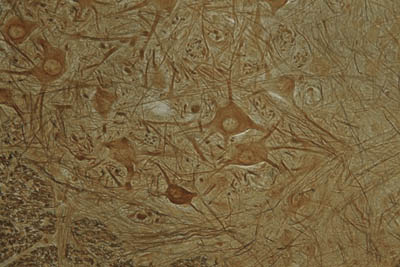
12. Golgi Apparatus. In the electron microscope,
this is seen as a system of parallel membranes arranged around the
nucleus. It is seen as black splotches
in the cytoplasm of large round nerve cells.
These nerve cells gave a large, light nucleus with a dark dot
(nucleolus).
________________________________________________________________________________________
13. Scalp. The skin consists of stratified squamous
epithelium, keratinized. The basal layer
is cuboidal, but becomes squamous in the upper layers. The cells die and form keratin, which are
layers of protein that slough off regularly.
Identify the hair follicles, sebaceous (oil) glands, sweat
glands and smooth muscles making up the arrector pili muscle.
________________________________________________________________________________________
14. Gut epithelium / Small
intestine (duodenum) simple columnar cells. Identify finger-like
processes (villi). The lining of these villi contains a simple columnar
epithelium with a striated or brush border (the electron
micrograph). The brush border can be
seen as a thin amorphous pink layer at the top of the epithelial cells. In most preparations some cells are different
from the simple columnar epithelium.
These appear as dark shadows or clear spots on the luminal surface of
the epithelium as you focus up and down.
These are the goblet cells, which are unicellular mucous
secreting cells. .
________________________________________________________________________________________
15. Parotid gland. Identify the secreting portion
and the ducts. Most of the gland is make
up of serous secreting alveoli (sacs of cells), which have characteristic
round, rather light, central nuclei and pink cytoplasm. The ducts look like necklaces, with the cells
being cuboidal, stratified cuboidal, to columnar. Identify the large ducts in the spaces
between lobules – some of these are stratified cuboidal.
Tuesday, November 15, 2016
Pre-lab Activity on Microscope, Cell, Mitosis and Stains
THIS IS TO BE DONE INDIVIDUALLY.
NAME: ___________________________
· Always begin examining microscope slides with which power objective? ____________
· The lens that is within the eyepiece of the light microscope is called the: ____________
·What must be done to a specimen to increase the contrast of the structures viewed? ____________
· The wheel under the stage that adjusts the amount of light is called the: ____________
· At high power, always use which adjustment knob to focus the image? ____________
· To focus a specimen, it is best to start with which objective: ____________
· Which objective provides the greatest depth of field? ____________
·The scanning, low, and high power objectives are mounted on the: ____________
If the ocular of a microscope is 10X and the objective is set at 100X, then what is the total magnification of the microscope? ____________
If the ocular of a microscope is 10X and the objective is set at 100X, then what is the total magnification of the microscope? ____________
1. This ____ (organelle) functions in cellular respiration.
2. The ____ (organelle) functions to package and deliver proteins.
3. Cell organelles are located within the ____ of the cell.
4. The endoplasmic reticulum functions to____________________
5. Genetic material is contained within the ___ of the cell.
6. This organelle is responsible for destroying worn-out cell parts___
7. The _____ controls what enters and leaves the cell.
8. The rough endoplasmic reticulum has ____ located on it.
9. Located within the nucleus, it is responsible for producing ribosomes:
10. Which structure is directly responsible for the formation of proteins within the cell.
Pre-test on Mitosis
· What is mitosis?
· Beginning with mitosis, what are the other parts of the cell cycle in order in which they occur?
· What occurs in the cell during the G2 phase?
· Beginning with interphase, what are the phases of mitosis in order in which they occur?
· In which phase do the chromosomes align on a plane in the center of the cell?
· What occurs during anaphase?
· What is the part of the chromosome that joins the two chromatids?
· What is the structure in cells that divides the cytoplasm into two cells.
· What is a blastula?
· What is the term that describes a nucleus with two of each type of chromosome?
A quiz on this assignment will be given on Friday. DO YOUR HONEST BEST!
Alkaline Phosphatase
Bielschowsky Stain


 Neurofibrillary Tangles associated with Alzheimer's
Cajal Stain
Congo Red
Giemsa Stain

STAINS (Study in advance)
Aldehyde Fuchsin- Used to stain pancreatic islet beta cell granules.
- This stains elastic fibers purple/black.

Alician Blue
- Stain mucins and mucosubstances blue.
- Copper in the histology stain is what ultimately is responsible for the blue color.
- Nuclei will stain pink/red.
- Cytoplasm stains a lighter pink.
- Mucins stain blue.


Alkaline Phosphatase
- Stain endothelial cells.
- Sites of alkaline phophatase activity will appear red.
- Nuclei will stain blue.
Bielschowsky Stain
- Silver is used in this histology staining process.
- This histology stain shows reticular fibers.
- This histology stain is also used for showing neurofibrillary tangles and senile plaques.
- Neurofibrillary tangles and senile plaques will stain black.
Neurofibrillary Tangles associated with Alzheimer'sCajal Stain

- This histology stain is used on nervous tissue
Congo Red
Congo red histology stain is used to stain amyloid.
- Amyloid will stain orange/red.
Giemsa Stain
- This is a histology stain for peripheral blood smears and bone marrow.
- It is also used to visualize parasites and malaria.
- This is a Romanowski type stain.
- Methylene blue and eosin are used.
- Erythrocytes stain pink/red.
- Platelets and leukocytes stain blue.
This micrograph depicts the appearance of a well-stained slide using the Giemsa staining technique.
Note that the acidic components of the cellular constituents such as the cytoplasm and chromatin, pick up the basic methylene blue azure compliments of the Giemsa stain, which reveals the characteristic blue
coloration of this stain.
Golgi Stain
Periodic Acid-Schiff (PAS)


Note that the acidic components of the cellular constituents such as the cytoplasm and chromatin, pick up the basic methylene blue azure compliments of the Giemsa stain, which reveals the characteristic blue
coloration of this stain.
Golgi Stain

- This histology stain will stain neurons.
H&E

- This is a standard histology stain.
- "H&E" stand for hematoxylin and eosin.
- Hematoxylin and eosin stain is used for routine tissue preparation frequently.
- This is the most often used combination in the histology lab for general purpose staining.
- Hematoxylin can be thought of as a basic dye.
- It binds to acidic structures, staining them blue to purple.
- It will bind and stain nucleic acids.
- Therefore, the nucleus stains blue.
- Eosin is an acid aniline dye.
- It will bind to and stain basic structures (or negatively charged structures), such as cationic amino groups on proteins.
- It stains them pink.
- Cytoplasm, muscle, connective tissue, colloid, red blood cells and decalcified bone matrix all stain pink to pink/orange/red with eosin.
- With an H&E stain, mucus and cartilage will stain a light blue color.
Iron Hematoxylin
- 
- stain nuclei bluish/black
- one of a number of stains that allow one to make a permanent stained slide for detecting and quantitating parasitic organisms
Oil Red O

- This is a histology stain used for lipids.
- Lipids will stain red.
- Nuclei will stain blue/black
Papanicolaou Stain
- This histology stain is used mainly on exfoliated cytological specimens.
- Cells in smear preparations can be stained with Pap staining.
- Gynecological smears (Pap smears), sputum, urine, cerebrospinal fluid, abdominal fluid, pleural fluid, synovial fluid, semminal fluid and fine needle aspiration samples can all be stained with a Pap stain.
- This staining technique involving five dyes in three solutions.
Periodic Acid-Schiff (PAS)
- This histology stain is particularly useful for staining glycogen and other carbohydrates, but is useful for many things.
- It is often used to show glomeruli, basement membranes, and glycogen in the liver.
- PAS stains glycogen, mucin, mucoprotein, and glycoproteins magenta.
- The nuclei will stain blue.
- Collagen will stain pink.
Prussian Blue

- used to stain iron (ferric iron and ferritin).
- demonstrates the blue granules of hemosiderin in hepatocytes and Kupffer cells.- Hemochromatosis
Safranin O

- This histology stain will stain mucin, cartilage and mast cells.
- It stains them orange/red.
- Safranin O is sometimes used as a counterstain.
Sudan Stains


- Sudan histology stains are used for staining of lipids and phospholipids.
- Examples of such histology stains are sudan black, sudan IV, and oil red O.
Subscribe to:
Comments (Atom)