Monday, February 13, 2017

Additional Reviewer Pointers

·         A muscle impulse travels deep into the muscle fiber along T-tubule membranes
·         A thick, fibrous connective tissue cord that attaches a muscle to a bone is a tendon
·         A typical bone has   the following major sets of blood vessels: nutrient;  metaphyseal and periosteal
·         Abundant molecules of hemoglobin give erythrocytes both their color and their ability to transport oxygen.
·         Adipose connective tissue is most similar to areolar, but is unique among connective tissues in having relatively little extracellular matrix.
·         Although they typically constitute 20-25% of all leukocytes, most lymphocytes reside in lymphatic tissues rather than blood.
·         Areolar  tissue is typically most closely associated with epithelia
·         At each end of a long bone is an expanded, knobby region called the epiphysis
·         Axon regeneration in the PNS involves neurolemmocytes in a process known as Wallerian degeneration
·         Cardiac muscle: occurs only in the myocardium; is both striated and involuntary; contains intercalated discs
·         Cartilage is composed of cells called chrondrocytes and may be surrounded by a covering called perichondrium
·         Clots formed by platelets and blood proteins protects the body against blood loss
·         Collectively, glial cells do all of the following:  protect and help nourish neurons; guide young migrating neurons during development; provide a supporting framework for all nervous tissue
·         Connective tissues perform all of the following functions: establishing the body's structural framework; defending the body from pathogens; transporting fluids and storing energy reserves
·         Dense connective tissue has the fewest cells per unit volume
·         Dense irregular connective tissue that forms a supporting layer around cartilage is called perichondrium
·         Dietary factors necessary for normal bone growth should include Vit D
·         Due to the fusion of embryonic myoblasts, skeletal muscle fibers get multiple nuclei
·         Embryonic connective tissues include mesenchyme and Wharton's jelly
·         Eosinophils phagocytize allergens and chemically attack parasitic worms               
·         Epithelial tissue  develops embryonically from all three primary germ layers
·         Erythrocytes are normally produced at the rate of about 3 million/sec.
·         Erythropoiesis occurs in red bone marrow
·         Fibroblast is the predominant cell type in areolar connective tissue  
·         Fibrocartilage is found in intervertebral discs and pubic symphysis           
·         Formed elements of blood: erythrocytes, leukocytes, and platelets
·         Functions of blood : transportation of oxygen and carbon dioxide; regulation of body temperature; defense against infection
·         Functions of bone:  storage of mineral and energy reserves; production of blood cells; support and protection
·         Hemopoiesis is the process of blood cell production
·         Hyaline cartilage, articular ends of long bones;  fibrocartilage, intervertebral discs and menisci; elastic cartilage, epiglottis and external ear
·         In neurons, the abundant free and bound ribosomes are collectively called the chromatophilic substance
·         In skeletal muscle fibers,   calcium ions occur at varying concentrations at: in the terminal cisternae of the sarcoplasmic reticulum; throughout the sarcoplasm; in regions where the thin and thick filaments overlap
·         Interstitial growth of a bone occurs in the epiphyseal plate
·         Intramembranous ossification produces the flat bones of the skull, some of the facial bones, the mandible, and the central part of the clavicle.
·         Leukocytes are divided into two classes based on the presence or absence of visible organelles specific granules
·         Leukocytes help initiate an immune response and defend the body against pathogens
·         Ligament is a band of dense regular connective tissue that attaches two bones?
·         Ligaments may be composed of either elastic or dense regular connective tissue
·         Lymphocytes that manage and direct an immune response, in some cases directly attacking foreign cells and virus-infected cells, are classified as T-lymphocytes
·         Mesenchyme: gives rise to all other connective tissues; is the first connective tissue to emerge in the embryo; persists as stem cells in some adult connective tissues
·         Monocytes are agranulocytes that leave the circulation after a few days to become macrophages
·         Neurons with numerous dendrites and a single axon are structurally classified as multipolar neurons
·         Neutrophils: most numerous type of leukocyte in the blood; kill bacteria by secreting an enzyme called lysozyme;  leave the blood to phagocytize pathogens in tissue spaces
·         Oligodendrocytes: large, bulbous cells with slender cytoplasmic extensions
·         Perforating canals and circumferential lamellae are components of compact bone, but not of an osteon
·         Physical conditioning can effectively decrease the proportion of fast muscle fibers?
·         Platelets: irregular, membrane-enclosed cellular fragments; less than one-fourth the size of an erythrocyte; normally about 120,000-300,000 per µl of adult blood                     
·         RBC: they transport oxygen from the lungs to body tissues; their plasma membrane contains many surface antigens; when mature, they have no nucleus or other organelles
·         Reflecting their different needs, fast muscle fibers contain large glycogen reserves whereas slow fibers contain the oxygen-carrying pigment myoglobin
·         Sarcoplasmic reticula extend into the sarcoplasm as a network of deep invaginations of the sarcolemma
·         Satellite cells are unfused myoblasts
·         Sensory nerves innervate  the bone matrix and marrow cavity and the periosteum and endosteum
·         Skeletal muscle : consists of long, cylindrical cells with multiple nuclei located at the periphery of the cell
·         Spongy bone contains slightly larger osteons than compact bone
·         Stacks of erythrocytes called rouleaux can pass through blood vessels barely larger than the diameter of a single erythrocyte.
·         Structural categories of neurons include  unipolar, bipolar, and multipolar
·         Synaptic knob describes the expanded tip of an axon at a neuromuscular junction
·         Synaptic knobs are to axons as motor end plates are to sarcolemmae, in that both are structural modifications involved in transmitting electrochemical signals across the synaptic cleft.
·         The bundle of dense regular connective tissue that attaches a skeletal muscle to bone is called a tendon.
·         The cell body of a mature neuron does not contain a centriole.
·         The continual process of producing new formed elements of all kinds is called hemopoiesis
·         The cytoplasm in a neuron cell body (or sometimes the entire cell body) is called the perikaryon
·         The endosteum: covers all internal surfaces of a bone, such as the medullary cavity; active during bone growth, repair, and remodelling; is an incomplete cellular membrane
·         The largest and most abundant of the CNS glial cells, astrocytes help form the blood-brain barrier.
·         The layer of dense irregular connective tissue that surrounds the entire skeletal muscle is the perimysium
·         The major functions of loose connective tissue include: occupying spaces between organs and supporting epithelia; supporting and surrounding blood vessels and nerves; organs, storing lipids, and facilitating diffusion
·         The minute passageways in the bony matrix that allow osteocytes to communicate with each other are called canaliculi
·         The slightly expanded tips of telodendria are called synaptic knobs
·         The three types of granulocytes, named according to how their granules stain: neutrophils, eosinophils, and basophils
·         The two primary components of whole blood are plasma and formed elements
·         The word root glia, as in "glial cells," most nearly means glue
·         Three basic components of connective tissue: Cells, protein fibers, and ground substance
·         Through the process of endochondral ossification, a fetal hyaline cartilage model transforms into bone.
·         Tiny, seed-shaped bones along the tendons of some muscles are classified as sesamoid bones or short bones
·         Two classes of cells are found in connective tissue proper:P resident cells and wandering cells
·         Types of skeletal muscle fibers include: fast; intermediate; slow
·         Wandering cells that may occur in connective tissue proper: mast cells, neutrophils, and free macrophages

·         Yellow bone marrow contains a large proportion of fatty tissue


Study also the previous reviewer posted 2 weeks ago.

Good Luck!

Tuesday, February 7, 2017

Pre-midterm exam Reviewer Pointers

Connective Tissue Generalities
  • Of the four basic tissue types (epithelium, connective tissue, muscle and nervous tissue), connective tissue is the most diverse. 
    • Blood, bone, tendon, and intervertebral discs are all composed of connective tissue. 
    • The myometrium is the muscular layer of the uterus. 
    • Thus, the myometrium is composed of muscle tissue.
  • There are three types of fibers found in connective tissue: collagen fibers, elastic fibers, and reticular fibers. 
    • Collagen fibers are the most abundant fiber type in connective tissue.
  • Purkinje fibers are seen in the heart. 
    • Purkinje fibers are specialized muscle fibers.
  • Myofibroblasts contain properties of both fibroblasts and smooth muscle cells.
  • Fibroblasts, histiocytes, plasma cells, and mast cells are routinely seen in loose connective tissue. 
    • Fibroblasts produce collagen. 
    • The fibroblast also produces the ground substance in connective tissue. 
    • Myofibroblasts contain properties of both fibroblasts and smooth muscle cells. 
    • The histiocyte is a connective tissue macrophage. 
    • Plasma cells are derived from B lymphocytes. 
    • Mast cells secrete histamine. 
    • Mast cells also secrete heparin, SRS-A (slow reacting substance of anaphylaxis, ECF-A (eosinophilic chemotactic factor of anaphylaxis.
  • Macrophages are mononuclear phagocytes. 
    • Many tissues have resident (fixed) macrophages. 
    • Fixed macrophages are given a unique name, depending on the tissue that they are located in. 
    • Kupffer cells are the hepatic macrophages. 
    • Histiocytes are macrophages seen in connective tissue. 
    • Dust cells are alveolar macrophage found in the respiratory tract. 
    • Langerhans cells are macrophages seen in the skin. 
    • Microglia are the central nervous system macrophages.
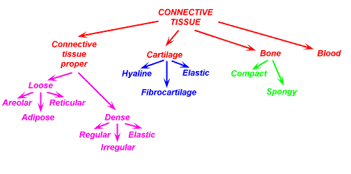
  • Connective tissue can be sub-classified into connective tissue proper, specialized connective tissue and embryonic connective tissue.
  • Connective tissue proper consists of loose irregular connective tissue and dense connective tissue (regular and irregular).
  • Specialized connective tissue includes cartilage, bone, adipose tissue, blood and hemopoietic tissue, and lymphatic tissue.
  • Embryonic connective tissue includes mesenchyme and mucous connective tissue.
  • Connective tissue can be sub-classified into connective tissue proper, specialized connective tissue and embryonic connective tissue.
  • Connective tissue proper consists of loose irregular connective tissue and dense connective tissue (regular and irregular).
  • Specialized connective tissue includes cartilage, bone, adipose tissue, blood and hemopoietic tissue, and lymphatic tissue.
  • Embryonic connective tissue includes mesenchyme and mucous connective tissue.
  • Mesenchyme is embryonic connective tissue. 
    • It is an undifferentiated tissue found in the embryo. 
    • Mucous connective tissue is a type of embryonic connective tissue; it is a subset of mesenchyme. 
    • Wharton's jelly is mucous connective tissue. 
    • Loose irregular connective tissue is areolar tissue. 
    • Dense irregular connective tissue is seen in the dermis. 
    • Dense regular connective tissue comprises tendons and ligaments.
  • Brown adipose tissue is multilocular adipose tissue. 
    • This is present during fetal development and then decreases after birth.
  • White adipose tissue is unilocular adipose tissue. 
    • This type of tissue persists into adulthood.
  • A peripheral blood smear would be best visualized with Wright's stain.
    •  Hematoxylin and eosin stain is the most commonly used tissue stain for routine histological examination. 
    • Lipids are best displayed with a sudan stain. 
    • Silver impregnation, such as with a reticular stain, can be used to visualize reticular fibers. 
    • Collagen fibers can be differentiated from other fibers by staining with Masson's trichrome stain.
Blood 

  • There are four basic tissue types: epithelium, connective tissue, muscle and nervous tissue. 
    • Connective tissue is the most diverse. 
    • Blood is considered a type of connective tissue.
  • Blood is composed of the formed elements and plasma. 
    • Plasma is the liquid portion of blood. 
    • The formed elements consist of the cells and cell fragments. 
    • The erythrocytes (red blood cells), platelets, and leukocytes (white blood cells) are all considered formed elements.
  • Erythrocytes do not contain a nucleus. 
    • During their development within the bone marrow, they have a nucleus. 
    • However, prior to their entering into the blood stream, the nucleus is ejected.
    • Platelets are cell fragments. 
    • Platelets also do not contain a nucleus. 
    • They are fragments from megakaryocytes within the bone marrow. 
    • Leukocytes, which include monocytes, are cells with a nucleus.
  • Platelets are cell fragments. 
    • They are fragments from megakaryocytes within the bone marrow.

  • Neutrophils are the most abundant leukocyte.
  • Leukocytes are the white blood cells.
    • The order of frequency of the leukocytes is: neutrophil, lymphocyte, monocyte, eosinophil, basophil. The order of frequency of the leukocytes can be remembered by the mnemonic: "Never Let Monkeys Eat Bananas".
  • The granulocytes are named because of the presence of visible cytoplasmic granules. 
    • The granulocytes consist of neutrophils, eosinophils, and basophils. 
  • "PMN" is an abbreviation for polymorphonuclear neutrophil. 
    • Thus, it is a neutrophil, which is a granulocyte. 
  • The non-granulocytes are lymphocytes and monocytes.

Muscle Tissue
  • The fundamental property of muscle tissue is that it is specialized for contraction.
  • The outer connective tissue covering of a muscle is the epimysium. 
  • Within the muscle, there are subdivisions called fascicles. 
  • The perimysium surrounds these muscle fascicles. 
    • The perimysium is the connective tissue sheath which surrounds muscle fascicles.
    • The periosteum is the connective tissue covering of a bone.
    • The perichondrium is the connective tissue which surrounds cartilage.
    • The perineurium is the covering of nerve fascicles.
    • The endosteum is the lining of the inner bone (the side which abuts the medullary cavity).
    • The endomysium is the covering around an individual muscle fiber. 

  • The sarcolemma is the plasma membrane of a muscle cell. 
  • The sarcoplasm is the cytoplasm of a muscle cell.
  • Myofilaments are the contractile protein within a muscle cell. 
  • On a cross section of a muscle, each thick filament is surrounded by 6 thin filaments.
  • The myofilaments are actin and myosin. The thin filaments are actin and the thick filaments are myosin. 
  • The muscle cell is a muscle fiber. The term "muscle cell" and "muscle fiber" are synonymous. 
  • A myofibril is a longitudinal bundle of myofilaments within a muscle cell. 
  • Myocardium is the muscular layer of the heart. 
  • Thus, the myocardium is composed of cardiac muscle.
  • There are two chief categories of muscle: striated and non striated muscle (smooth muscle).
    • Striated muscle can be sub-categorized into cardiac muscle and skeletal muscle. The tongue, biceps muscle, and upper esophagus are made of striated muscle. The heart is composed of cardiac muscle.
    • Non striated muscle is also called smooth muscle. Smooth muscle is involuntary muscle. It is found in viscera and blood vessels.
  • Smooth muscle is composed of spindle shaped cells.
  • Skeletal muscle is multinucleated. The nuclei are seen on the periphery. Occasionally, cardiac muscle is bi-nucleated.
  • Skeletal muscle fibers can be classified as red fibers, white fibers or intermediate fibers. 
    • Red fibers are smaller in diameter; white fibers are larger in diameter. 
    • Red fibers have more mitochondria than white fibers. 
    • Red fibers make up slow-twitch muscle; white fibers make up fast-twitch muscle. 
    • Red fibers are more resistant to fatigue than are white fibers. 
    • Red fibers have more myoglobin (oxygen binding pigment) than white fibers. 
    • White fibers store glycogen and use anaerobic metabolism.
    • Red fiber and slow twitch muscle is for endurance.
    • White fiber and fast twitch muscle is for a burst of power.
    • The A band is the darker staining band. 
    • The I band is the light band. 
    • The I band is made of thin filaments. 
    • The Z line runs through the I band. 
    • The H band bisects the A band. The M line runs through the H band.
    • A sarcomere is the segment that runs from Z line to Z line.
  • Both skeletal muscle and cardiac muscle have visible striations. Collectively, skeletal muscle and cardiac muscle are classified as "striated muscle".
  • A single motor neuron and the aggregation of muscle fibers innervated by that single neuron is called the motor unit. 
  • A motor neuron is a neuron which innervates a muscle cell. 
  • The point of contact where a neuron contacts a muscle is the motor end plate. 
  • A neuromuscular spindle is a receptor which is sensitive to stretching of the muscle. 
  • A neurotransmitter is the chemical released by a nerve at a synapse.
BoneTissue
  • Of the four basic tissue types (epithelium, connective tissue, muscle and nervous tissue), connective tissue is the most diverse. 
    • Bone is a type of connective tissue.
  • "Bone" can refer to either a type of tissue or to the organ. 
    • As a tissue, bone tissue is a type of connective tissue. 
    • When referring to a bone (the organ), there will be several types of tissue present.
  • Compact bone is also called dense bone. 
    • Compact bone has the Haversian system.
  • Immature bone is woven bone. 
    • It is nonlamellar bone or bundle bone.
  • Spongy bone is also referred to as cancellous bone. 
    • The mineralized tissue is seen as spicules. 
    • Marrow spaces are also present.
  • An osteoclast is a multinucleated cell involved in the degradation of bone. 
    • It is a bone resorbing cell.
  • An osteon is the cylindrical structure with bone. 
    • An osteon is also called a Haversian system.
  • The mature bone cell is called an osteocyte. 
    • It sits in a space, called a lacuna.
  • An osteoblast is an immature bone cell. 
    • The osteoblast is the bone forming cell.
  • Osteoid is unmineralized bone matrix.
  • Canaliculi are the little tunnels within bone.
  • Sharpey's fibers are collagen fibers that extend into a bone at an angle.
  • Trabeculae are the spicules seen with spongy bone.
  • Tome's process is seen in teeth, this process is responsible for enamel production.
  • An osteocyte rests in a space called a lacuna.
  • The mature cell in cartilage is a chondrocyte. 
    • It rests in a lacunae surrounded by matrix. 
    • A chondroblast is an immature cartilage cell which produces the cartilaginous matrix. 
    • An osteocyte is a mature bone cell. 
    • An osteoclast is a bone cell which is involved in resorption of bone. 
    • A bone lining cell is a resting osteoblast.
  • Intramembranous bone formation is the process of bone formation where the bone is formed without a cartilage template. 
    • Endochondral bone formation is the process of bone formation where the bone is formed using a cartilage template.
  • Hyaline cartilage forms the epiphyseal growth plate.
  • The mature cell in cartilage is a chondrocyte. It rests in a lacunae surrounded by matrix. A chondroblast is an immature cartilage cell which produces the cartilaginous matrix. An osteocyte is a mature bone cell. An osteoclast is a bone cell which is involved in resorption of bone. A bone lining cell is a resting osteoblast.
  • Calcium is in a mineral structure in bone and tooth enamel called hydroxyapatite. 
    • The chemical formula is [Ca10 (PO4)6(OH)2]
  • Calcite crystals are calcium carbonate. 
    • The main component of limestone is calcite and seashells are made of calcite. 
      • However, calcite crystals are not found in man.
  • Tourmaline is a crystal found in nature. 
    • Pink-red tourmaline is called rubellite. 
    • Blue tourmaline is known as indicolite. 
    • Tourmaline crystals are not found in man, although sometimes they are found on the necks and fingers of women in the form of jewelry.
  • Red marrow contains active hematopoietic tissue. Yellow marrow is primarily fat.

Nervous Tissue:
  • Schwann cells are responsible for the myelination of neurons in the peripheral nervous system. 
    • Basket cells are a type of neuron seen in the cerebellum. 
    • A ganglion is a collection of nerve cell bodies outside of the CNS. 
    • Neuroglia are the supporting cells in the central nervous system 
    • Sometimes, neuroglia called glial cells or glia. 
    • Satellite cells are found in the ganglia of the peripheral nervous system.
  • The central nervous system consists of the brain and spinal cord. 
    • All other nervous system elements are considered to be in the peripheral nervous system. 
    • Thus the peripheral nervous system includes receptors, the brachial plexus, the sciatic nerve, and ganglia.
  • Most neurons in the body (over 99%) are multipolar.
  • Neurons can be classified based on the number of axons and dendrites stemming off of the cell body. 
    • A unipolar neuron has one process which branches off of it This process then immediately divides into two. Thus, a unipolar neuron is sometimes also called a pseudounipolar neuron. Sensory neurons are unipolar.
  • A bipolar neuron has two process that branch from it: an axon and a dendrite. 
    • Bipolar neurons are not very common and are found in some of the organs for special senses. 
    • Bipolar neurons are found in the retina, inner ear, and the region of the nose involved with smell.
  • Multipolar neurons have one axon and many (at least two) dendrites that branch off of it. 
    • Most neurons are multipolar. 
    • Motor neurons and interneurons are multipolar.
  • A ganglion is a collection of neuron cell bodies outside of the central nervous system. 
    • The cell body of a neuron is called a perikaryon or soma. 
    • An astrocyte is a supporting cell seen in the central nervous system. 
    • The basophilic clusters of ribosomes and rough endoplasmic seen in neuron cell bodies is called Nissl, Nissl bodies or Nissl substance. 
    • The terminal bouton is the end portion of an axon. 
    • It is also called an axon terminal or end bulb. 
    • The terminal bouton will be associated with another neuron in a synapse.
  • Macrophages are mononuclear phagocytes. 
    • Many tissues have resident (fixed) macrophages. 
    • Fixed macrophages are given a unique name, depending on the tissue that they are located in. Kupffer cells are the hepatic macrophages. 
    • Histiocytes are macrophages seen in connective tissue. 
    • Dust cells are alveolar macrophage found in the respiratory tract. 
    • Langerhans cells are macrophages seen in the skin. 
    • Microglia are the central nervous system macrophages.
  • Both motor neurons and interneurons are multipolar.
  • The meninges are composed of the three connective tissue covers which surround the brain and spinal cord. 
    • White matter is myelinated nerve fibers.
    • Gray matter is essentially neuron cell bodies and associated dendrites or unmyelinated axons. 
    • Nodes of Ranvier are the gaps that occur in the myelin sheath. 
    • Neurofilaments are a type of intermediate filaments seen in neurons which provide rigidity and tensile strength.
  • The meninges cover the brain and spinal cord. 
    • There are three: dura mater, arachnoid, and pia mater.
  • The dura mater is the outer meninx. 
    • The dura mater is composed of dense irregular connective tissue.
  • The arachnoid is made of a delicate spider web like connective tissue.
  • The pia mater is the innermost meninx. 
    • It covers the brain intimately.
  • A group of fibers traveling together to a destination is a tract. 
    • Islets of Langerhans are seen in the pancreas. 
    • Soma is another term for a neuron cell body. 
    • The outer part of the brain is gray matter called the cortex. 
    • The conglomerations of gray matter deep within the cerebrum and cerebellum are called nuclei.
  • Neuroglia are the supporting cells of the central nervous system. 
    • Sometimes, neuroglia called glial cells or glia. 
    • Astrocytes, ependymal cells, oligodendrocytes, and microglia are all neuroglia.
  • Of the neuroglia cells, astrocytes are the most abundant and the largest. 
    • These are star shaped cells involved in the blood brain barrier.
    • Ependymal cells line the ventricles and spinal canal.
    • Oligodendrocytes form myelin in the central nervous system.
    • Microglia are the central nervous system macrophages.
    • Schwann cells are seen in the peripheral nervous system and are not considered neuroglia. 

Tuesday, January 31, 2017

FEB. 1 Activity: Simulated Lab - Video Viewing Nervous Tissue

INDIVIDUALLY

1.Transcribe (Transcription means: extracting all the factual information from a lecture or discussion; notes taking.) the discussion as presented in the link below by creating an Atlas of your own. Screen Capture pictures must be labelled correctly.


2. Label the following by matching the specified parts with the list of terms that appear to the left of each picture/diagram:

3. Enumerate and describe the location and functions of the unique cells of the Nervous system.
Example: (Purkinje cells - Cerebellum)

TO BE SUBMITTED ONLINE TO: apbautista@ceu.edu.ph
Due before 1 pm today.
No need to go to the LAB today.

Tuesday, January 24, 2017

January 25. 2017 Activity

In groups of 5 members each, research for photomicrographs and or sketch of a cross section of the following: 
1.Structure of a typical motor neuron (dendrites, soma, axon, synaptic terminals) - Subtypes
2.Neuroglia. (a) Astrocyte. (b) Microglial cell. (c) Ependymal cells. (d) Oligodendrocyte. (e) Neuron with Schwann cells and satellite cells.*
3.Specialized synapses in skeletal muscle are called neuromuscular junctions. 

  1. Cranial n. (CN V) vs. Peripheral Nerve - Sciatic nerve  (x.s. and l.s.) Identify nerve fibers, myelin sheaths, fascicles, and endoneurium, perineurium, and epineurium sheaths.
  2. Cerebrum - unique cells; brain centers
  3. Cerebellum - - unique cells; brain centers
  4. Unique cells; brain centers; Brainstem – Midbrain; Pons Medulla 
  5. Spinal Cord
  6. Spinal ganglion 
  7. Longitudinal view of myelinated axon showing portions of adjacent Schwann cells and the node of Ranvier between them. 
  8. Non - myelinated axon.
  9. Illustrate (sketch) the process of Myelination of neuron
Finish this at the library up to 1:30 pm and submit to:
Hard copy, scan or take a picture; send to apbautista@ceu.edu.ph

Hard copy will be presented on Friday, pretest on Bones and Nervous Tissue will be given.

Tuesday, January 10, 2017

Connective Tissue

Of the four basic tissue types (epithelium, connective tissue, muscle and nervous tissue), connective tissue is the most diverse. Connective tissue can be found all over the body. Connective tissue cells are capable of reproducing, however not as fast as epithelial cells.  Embryologically, connective tissue develops from mesenchyme.

Function of Connective Tissue

Connective tissues connects and holds tissues and organs to one another, forms a framework, provides support, stores fat, transports things, protects, and aids in tissue repair.


Histology of Connective Tissue

Connective tissue consists of cells and extracellular fibers in a ground substance and tissue fluid. The cells within connective are not tightly packed together. On a histology slide, it can be seen that there is generally abundant extracellular space in connective tissue.

Within connective tissue, the cells and fibers are scattered within the ground substance. The ground substance is amorphous material composed of proteoglycans and glycosaminoglycans. Hyaluronic acid is a glycosaminoglycan. Dermatan sulfate, chondroitin sulfate, and keratan sulfate are also
glycosaminoglycans.

Most of the connective tissues in the body have a good vascular supply.


Cell Types in Connective Tissue

Numerous cell types are found in connective tissue. Fibroblasts, histiocytes, plasma cells, and mast cells are cell types that are found in loose connective tissue.

FibroblastRelated image

A fibroblast is a connective tissue cell derived from mesenchyme.   Fibroblasts produce collagen. The fibroblast also produces the ground substance in connective tissue.

HistiocyteImage result for Histiocyte

The histiocyte is a connective tissue macrophage.  Macrophages are mononuclear phagocytes, derived from a monocyte. A macrophage is a phagocytic tissue cell of the reticuloendothelial system that may be fixed or freely motile. Many tissues have resident (fixed) macrophages. Fixed macrophages are given a unique name, depending on the tissue that they are located in. In connective tissue, the fixed macrophage is a histiocyte. The function of histiocytes is for the protection of the body against infection and other harmful things.

Image result for Histiocyte

Plasma cells are derived from B lymphocytes.

Mast Cell

Image result for Mast Cell
A mast cell is a large cell seen in connective tissue.  A mast cell has basophilic granules containing histamine and heparin which mediate allergic reactions. Mast cells also secrete SRS-A (slow reacting substance of anaphylaxis and ECF-A (eosinophilic chemotactic factor of anaphylaxis).


Fibers in Connective Tissue

Image result for collagen fibersImage result for reticular  fibers
There are three types of fibers found in connective tissue: collagen fibers, elastic fibers, and reticular fibers. Collagen fibers are the most abundant fiber type in connective tissue.


Classification of Connective Tissue
Image result for connective tissue proper types

Connective tissue can be sub-classified into connective tissue proper, specialized connective tissue and embryonic connective tissue.

Connective Tissue Proper
Image result for connective tissue proper types

Connective tissue proper consists of loose irregular connective tissue and dense connective tissue. Dense connective tissue is subdivided into dense regular connective tissue and dense irregular connective tissue.

Related imageImage result for Dense regular connective tissue comprises tendons and ligaments.
Related image
Loose Irregular Connective Tissue
Related image
Loose irregular connective tissue is areolar tissue.

Dense Regular Connective Tissue
Related image
Dense regular connective tissue comprises tendons and ligaments.

Dense Irregular Connective Tissue

Dense irregular connective tissue is seen in the dermis.

Specialized Connective TissueImage result for Specialized Connective Tissue
Specialized connective tissue includes cartilage, bone, adipose tissue, blood and hemopoietic tissue, and lymphatic tissue.
Cartilage
Bone
Adipose Tissue


Brown adipose tissue is multilocular adipose tissue. Brown adipose tissue is present during fetal development and then decreases after birth. White adipose tissue is unilocular adipose tissue. White adipose tissue persists into adulthood.


Image result for brown vs white adipose tissue
Blood and Hemopoietic Tissue

Lymphatic Tissue

Embryonic Connective Tissue

Image result for embryonic connective tissue labeled
Embryonic connective tissue includes mesenchyme and mucous connective tissue.  Mesenchyme is embryonic connective tissue. It is an undifferentiated tissue found in the embryo. Mucous connective tissue is a type of embryonic connective tissue; it is a subset of mesenchyme. Wharton's jelly is mucous connective tissue.

Histotechniques

Image result for Collagen fibers stain green with Masson's trichrome stain
Collagen fibers stain green with Masson's trichrome stain. Collagen fibers can be differentiated from other fibers by staining with Masson's trichrome stain.  A trichrome stain is a mixture of three dyes.
Image result for Gomori's trichrome will stain connective tissue green, cytoplasm red, and nuclei gray blue black.
Gomori's trichrome will stain connective tissue green, cytoplasm red, and nuclei gray blue black.
Verhoeff Elastic stain stains elastic fibers blue/black. Collagen stains pink/red.

KEY POINTERS FOR REVIEW:
  • Of the four basic tissue types (epithelium, connective tissue, muscle and nervous tissue), connective tissue is the most diverse. Blood, bone, tendon, and intervertebral discs are all composed of connective tissue. The myometrium is the muscular layer of the uterus. Thus, the myometrium is composed of muscle tissue.
  • There is connective tissue in the heart. The blood in the heart, for example, is composed of connective tissue. The pericardium is also composed of connective tissue. However, the primary tissue composing the heart is cardiac muscle.
  • There are three types of fibers found in connective tissue: collagen fibers, elastic fibers, and reticular fibers. Collagen fibers are the most abundant fiber type in connective tissue.
  • Myofibroblasts contain properties of both fibroblasts and smooth muscle cells.
  • Fibroblasts, histiocytes, plasma cells, and mast cells are routinely seen in loose connective tissue. Fibroblasts produce collagen. The fibroblast also produces the ground substance in connective tissue. Myofibroblasts contain properties of both fibroblasts and smooth muscle cells. The histiocyte is a connective tissue macrophage. Plasma cells are derived from B lymphocytes. Mast cells secrete histamine. Mast cells also secrete heparin, SRS-A (slow reacting substance of anaphylaxis, ECF-A (eosinophilic chemotactic factor of anaphylaxis.
  • Macrophages are mononuclear phagocytes. Many tissues have resident (fixed) macrophages. Fixed macrophages are given a unique name, depending on the tissue that they are located in. Kupffer cells are the hepatic macrophages. Histiocytes are macrophages seen in connective tissue. Dust cells are alveolar macrophage found in the respiratory tract. Langerhans cells are macrophages seen in the skin. Microglia are the central nervous system macrophages.
  • Connective tissue can be sub-classified into connective tissue proper, specialized connective tissue and embryonic connective tissue.

  • Connective tissue proper consists of loose irregular connective tissue and dense connective tissue (regular and irregular).

  • Specialized connective tissue includes cartilage, bone, adipose tissue, blood and hemopoietic tissue, and lymphatic tissue.
  • Embryonic connective tissue includes mesenchyme and mucous connective tissue.
  • Mesenchyme is embryonic connective tissue. It is an undifferentiated tissue found in the embryo. Mucous connective tissue is a type of embryonic connective tissue; it is a subset of mesenchyme. Wharton's jelly is mucous connective tissue. Loose irregular connective tissue is areolar tissue. Dense irregular connective tissue is seen in the dermis. Dense regular connective tissue comprises tendons and ligaments.
  • Brown adipose tissue is multilocular adipose tissue. This is present during fetal development and then decreases after birth.

  • White adipose tissue is unilocular adipose tissue. This type of tissue persists into adulthood.

  • A peripheral blood smear would be best visualized with Wright's stain. Hematoxylin and eosin stain is the most commonly used tissue stain for routine histological examination. Lipids are best displayed with a sudan stain. Silver impregnation, such as with a reticular stain, can be used to visualize reticular fibers. Collagen fibers can be differentiated from other fibers by staining with Masson's trichrome stain.
  • Connective tissue develops from mesenchyme.
  • Within connective tissue, the cells and fibers are embedded in the ground substance. The ground substance is amorphous material. It is composed of proteoglycans and glycosaminoglycans. Hyaluronic acid and chondroitin sulfate are glycosaminoglycans.
  • Fibroblasts produce collagen. The fibroblast also produces the ground substance in connective tissue.
  • Plasma cells are derived from B lymphocytes.
  • Connective tissue consists of cells and extracellular fibers in a ground substance and tissue fluid. There is generally abundant extracellular space in connective tissue

Thursday, December 8, 2016

Connective tissue Prelim Exam Requirement:

INDIVIDUALLY Submit a transcription of this video on connective tissue: on December 16, 2016

Tuesday, December 6, 2016

Connective tissue


1. Remember that all connective tissue contains
  • -cells 
  • -fibers 
  • -collagen 
  • -elastic 
  • -reticular 
  • -ground substance (extracellular matrix) 
2. In photomicrographs, identify and give the location of all the types of connective tissue:
  • -fetal mesenchyme 
  • -mucoid connective tissue 
  • -loose, areolar fibrous CT (e.g., mesentery) 
  • -dense, irregular fibrous CT 
  • -dense, regular fibrous CT 
  • -elastic CT 
  • -reticular CT 
  • -adipose CT 
  • -white (unilocular) fat 
  • -brown (multilocular) fat 
3. Give the basic functions of the two major resident cells of connective tissue:
  • -fibroblasts 
  • -adipocytes 
4. Explain the role of the transient cells of connective tissue:
  • -macrophage 
  • -mast cell 
  • -plasma cell 
  • -leukocytes 
5. Define the terms “agyrophilia and metachromasia.”

6. List the general functions of connective tissues 

7. Name the germ layer(s) from which connective tissue cells derive and the embryonic tissues containing undifferentiated connective tissue cells  
8. List the two major classes of macromolecules that constitute ground substance
9. Describe adipose tissue as a connective tissue in terms of its cells, fibers, and ground substance.
10. Compare the three types of cartilage in terms of:
  1. Type, amount, and arrangement of cells, fibers, and ground substance
  2. Location in the body 
  3. Histogenesis 
  4. Function
11. Compare osteocytes with osteoblasts in terms of their shape, filopodia, amount of RER, location, and rate of matrix synthesis.

SUBMIT ANSWERS PER GROUPS OF 8 MEMBERS EACH TO apbautista@ceu.edu.ph before 2 pm today (12-7-16)
Additional points will be given to:
1st - 20 pts to CP and 5 pts to the quiz on Friday
2nd - 15 pts
3rd - 10 points

Post lab discussion will be on Friday (12-9-16) and a short quiz will be given on connective tissue and epithelial tissue (30 pts)

PRELIM EXAM WILL BE ON FRIDAY NEXT WEEK (12-16-16)